AI methods for blood vessel segmentation presented at international conferences
The team from the Medical Informatics research department at RISC Software GmbH has presented three groundbreaking AI methods for blood vessel segmentation at two international conferences. This work enables more precise diagnoses of vascular diseases.
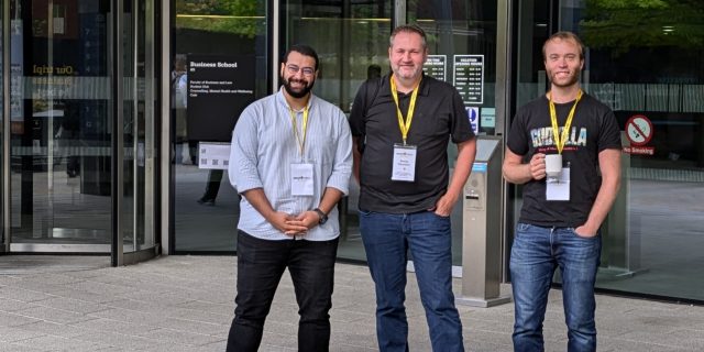
Ahmed AlShenoudy, Stefan Thumfart and Bertram Sabrowsky-Hirsch at the MIUA 2024 in Manchester
Success at the MIDL 2024 conference in Paris
Employees from the Medical Informatics department at RISC Software GmbH presented a poster on the segmentation of cerebral arteries at the renowned Medical Imaging with Deep Learning (MIDL) Conference 2024 in Paris. This method makes it possible to analyze structural MRI data more efficiently and improves the diagnosis of vascular diseases in the brain. The results can be read in the paper “Brain Artery Segmentation for Structural MRI”. The MIDL paper presents a combined approach of registration and segmentation (i.e. both MIUA papers – see below), which improves the former approach and enables automatic extension with additional training datasets. More details and the full paper can be found here.
Presentations at the MIUA 2024 in Manchester
In addition, the research department presented two further research papers at the Medical Image Understanding and Analysis (MIUA) Conference 2024 in Manchester. The presentation “Towards Segmenting Cerebral Arteries from Structural MRI” highlighted advanced techniques for segmenting cerebral arteries. This work shows how AI can improve the process of analyzing blood vessels in MRI images. The paper lays the groundwork, the novelty aspect lies in the segmentation of structural MRIs (instead of usual angiography images). Further information and access to the paper can be found here.
A second presentation dealt with the “Robust Multi-Modal Registration of Cerebral Vasculature”. This paper describes a method for the precise registration of blood vessels across different image modalities. This technique is an essential step towards better analysis and treatment of vascular diseases. The paper is available here.
GitHub publication
The team has also published the models and methods developed on GitHub. Researchers and developers can view them on GitHub and use them for their own projects.
Added value when combining image data
The added value of the methods in clinical application results in the diagnosis from combined multimodal image data (angiography and structural MRIs) as these can be automatically superimposed. This is particularly helpful when the surrounding brain tissue is of interest in addition to the vessel geometry (hemorrhage, localization in fissures, etc.). It is also a first step towards the diagnosis of vascular disease as a secondary indication (from structural MRIs, without angiography), even if the accuracy is currently not sufficient for this.
Papers
Sabrowsky-Hirsch, B., AlShenoudy, A., Thumfart, S., Giretzlehner, M., & Scharinger, J. (2024). Brain Artery Segmentation for Structural MRI. In Medical Imaging with Deep Learning. OpenReview. Link.
Alshenoudy, A., Sabrowsky-Hirsch, B., Scharinger, J., Thumfart, S., Giretzlehner, M.. (2024). Towards Segmenting Cerebral Arteries from Structural MRI. 28th UK Conference on Medical Image Understanding and Analysis (MIUA 2024). Springer Nature. Link.
Sabrowsky-Hirsch, B., Alshenoudy, A., Scharinger, J., Gmeiner, M., Thumfart, S., Giretzlehner, M.. (2024). Robust Multi-Modal Registration of Cerebral Vasculature. 28th UK Conference on Medical Image Understanding and Analysis (MIUA 2024). Springer Nature. Link.
MIDL 2024 in Paris (Medical Imaging with Deep Learning (MIDL) Conference 2024)
Bertram Sabrowsky-Hirsch auf der MIDL 2024 in Paris (Medical Imaging with Deep Learning (MIDL) Conference 2024)
MIUA 2024 in Manchester (Medical Image Understanding and Analysis (MIUA) Conference 2024)
MIUA 2024 in Manchester (Medical Image Understanding and Analysis (MIUA) Conference 2024)
MIUA 2024 in Manchester (Medical Image Understanding and Analysis (MIUA) Conference 2024)
Ahmed AlShenoudy und Bertram Sabrowsky-Hirsch auf der MIUA 2024 in Manchester (Medical Image Understanding and Analysis (MIUA) Conference 2024)
Ahmed AlShenoudy, Stefan Thumfart und Bertram Sabrowsky-Hirsch auf der MIUA 2024 in Manchester (Medical Image Understanding and Analysis (MIUA) Conference 2024)
Ahmed AlShenoudy, Stefan Thumfart and Bertram Sabrowsky-Hirsch at the MIUA 2024 in Manchester (Medical Image Understanding and Analysis (MIUA) Conference 2024)