Blood flow simulation in the brain on the basis of medical image data.
The goal of the research project MEDVIS 3D, carried out in cooperation with Kepler University Hospital Linz (Med Campus III, formerly AKH Linz, and Neuromed Campus, formerly LNK Wagner-Jauregg), was the development of a universal software tool for the easy and fast reconstruction of aneurysms from medical image data (MR, CT, etc.). Based on the intensity data of these modalities, the MEDVIS 3D software reconstructs and directly visualizes the acquired volume in 3D. Furthermore, the system marks areas that contain pathologically dilated vessels (aneurysms) and calculates various measures such as diameters or volume. Physicians can define inlet planes through direct interaction, and the system automatically reconstructs the vessel pathway between them. Thus, the software not only supports the diagnosis of aneurysms but also enables a quantitative assessment of therapy progress.
From Geometry to Blood Flow Simulation
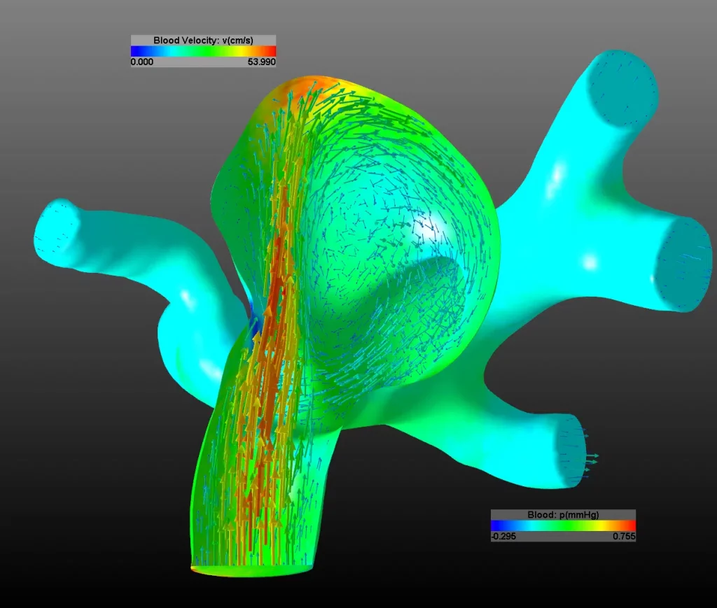
Reconstructed 3D geometry data can be integrated into a computer simulation model. In particular, the Finite Element Method (FEM) allows the system to simulate blood flow in the brain. The complex numerical solvers are accelerated by the Algebraic MultiGrid method (AMG), High Performance Computing (HPC) and modern GPU programming. Depending on the vessel structure’s complexity, a simulation run may take only a few minutes or extend over several hours. The MEDVIS 3D system calculates velocity and pressure fields, as well as surface stresses at the vessel wall. In addition, it can determine the displacement of the vessel wall caused by the blood pulse.
Clinical Impact
From these simulation results, physicians can derive both the risk of rupture of the dilated vessel (hemorrhage) and appropriate therapy measures. The system was evaluated together with medical experts in clinical routine, and all results were stored in a central database. Consequently, this collaboration led to a deeper understanding of the mechanical processes involved in the development and growth of aneurysms. A clinically usable version of MEDVIS 3D has existed since 2013 and continues to support diagnosis and treatment planning.
This work was financed by research subsidies granted by the province of Upper Austria, GESPAG (now OÖG) and FFG.